Facile Synthesis of Oleic Acid Capped Iron Oxide Nanoparticles as a Hyperthermic Agent for Cancer Cells Lysis
International Journal of Nanoparticles and Nanotechnology
(ISSN: 2631-5084)
Volume 7, Issue 1
Research Article
DOI: 10.35840/2631-5084/5539

Article Formats
Facile Synthesis of Oleic Acid Capped Iron Oxide Nanoparticles as a Hyperthermic Agent for Cancer Cells Lysis
Table of Content
Figures
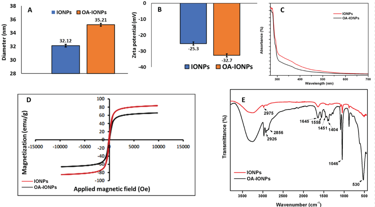
Figure 1: Analysis of non-coated...
Analysis of non-coated and OA-coated IONPs. A) DLS analysis showing the change in hydrodynamic diameter; B) Zeta potential analysis showing a decrease in surface charge; C) UV-vis spectrophotometry showing a decrease in absorbance intensity; D) VSM analysis showing the difference in superparamagnetic behaviour of IONPs and OA-IONPs and E) FT-IR spectroscopy confirming the shift in peaks of IONPs after OA coating.
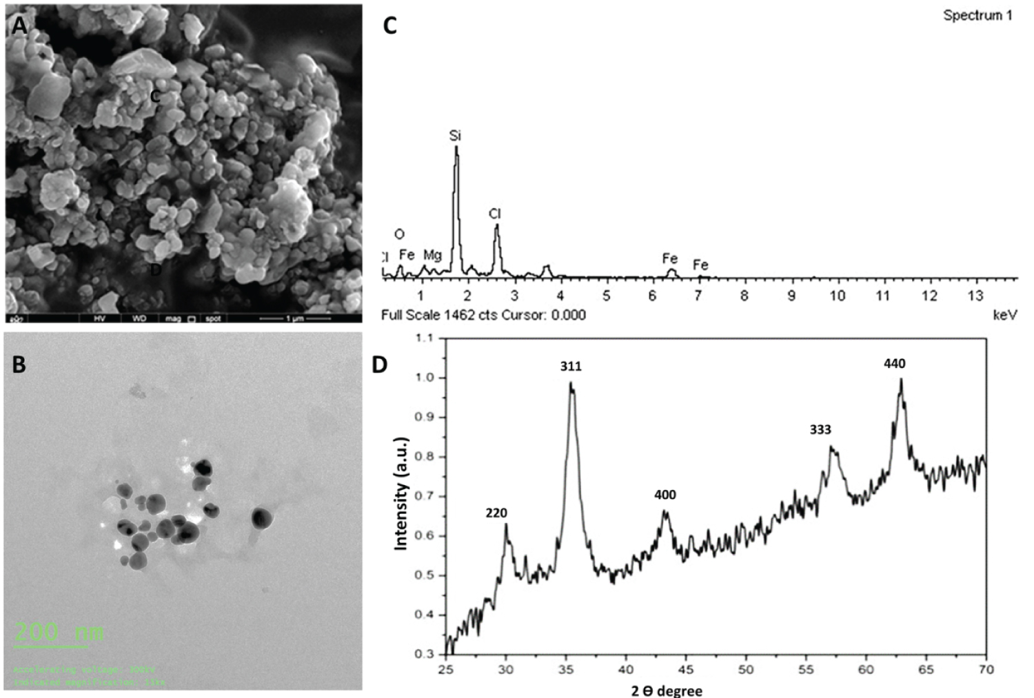
Figure 2: Morphological features and....
Morphological features and elemental analysis of OA-IONPs. A) FESEM image showing homogenous texture; B) TEM image showing spherical shaped nanoscale particles; C) EDX confirming the presence of Fe and other residual elements and D) XRD analysis showing characteristic diffraction peak of OA-IONPs.
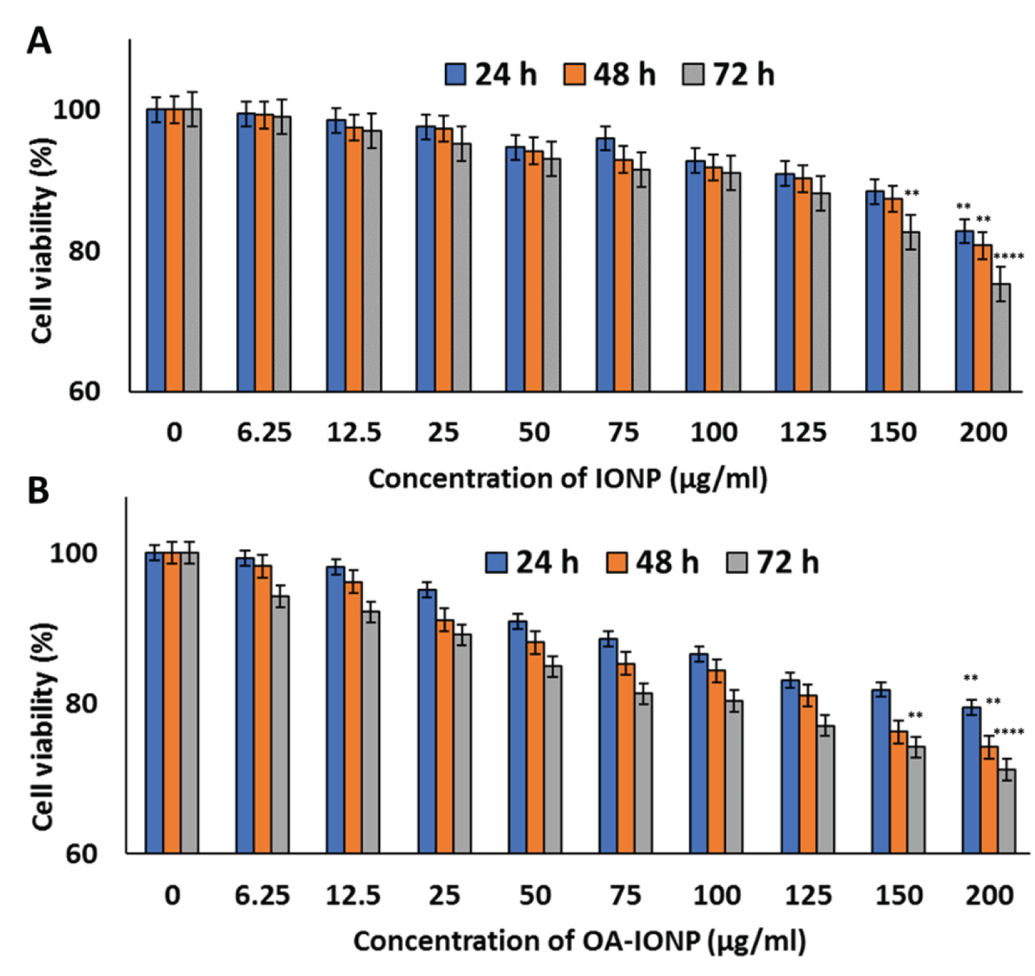
Figure 3: Viability study of MDA-MB-231....
Viability study of MDA-MB-231 cells on treatment with different concentrations of (A) IONPs and (B) OA-IONPs for 24, 48, and, 72 h, suggesting non-toxic behaviour of synthesized MNPs even at prolonged exposure and high dose. Data are expressed as mean ± standard deviation for n = 6 for both the sets.
*p < 0.05, **p < 0.005, ***p < 0.001, and, ****p < 0.0001 in comparison to control (untreated) cells.
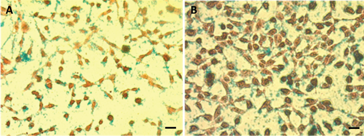
Figure 4: Prussian blue assay showing....
Prussian blue assay showing the presence of iron after treatment with OA-IONPs in MDA-MB-231 cells, in A) Control cells and B) OA-IONPs treated cells. A blue corona around cells depicts the presence of iron. Scale bar = 20 µm.
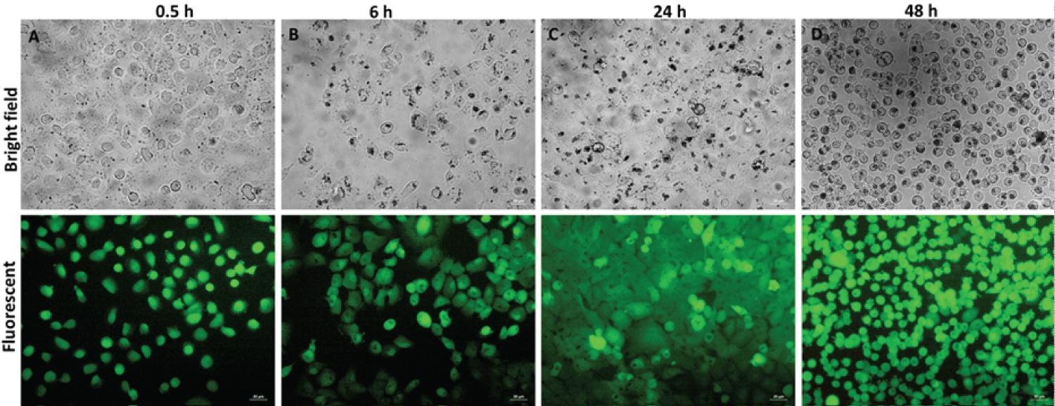
Figure 5: Bright-field and fluorescence....
Bright-field and fluorescence images of GFP-tagged MDA-MB-231 cells showing invasion of OA-IONPs with a dose of 100 µg/mL at different time intervals for 48 h, where black clusters represent the presence of OA-IONPs. Scale bar = 20 µm.
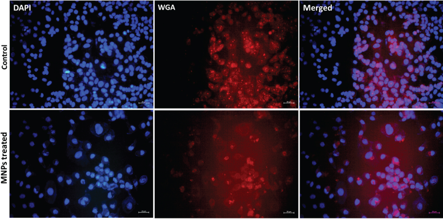
Figure 6: Fluorescence images of MDA-MB-231....
Fluorescence images of MDA-MB-231 cells after treatment with MNPs (100 µg/mL) at 48 h. DAPI (excitation/emission: 358⁄461 nm) as nucleus marker and WGA, Alexa Fluor 633 (excitation/emission: 632/647 nm) as cell membrane marker were used. Scale bar = 20 µm.
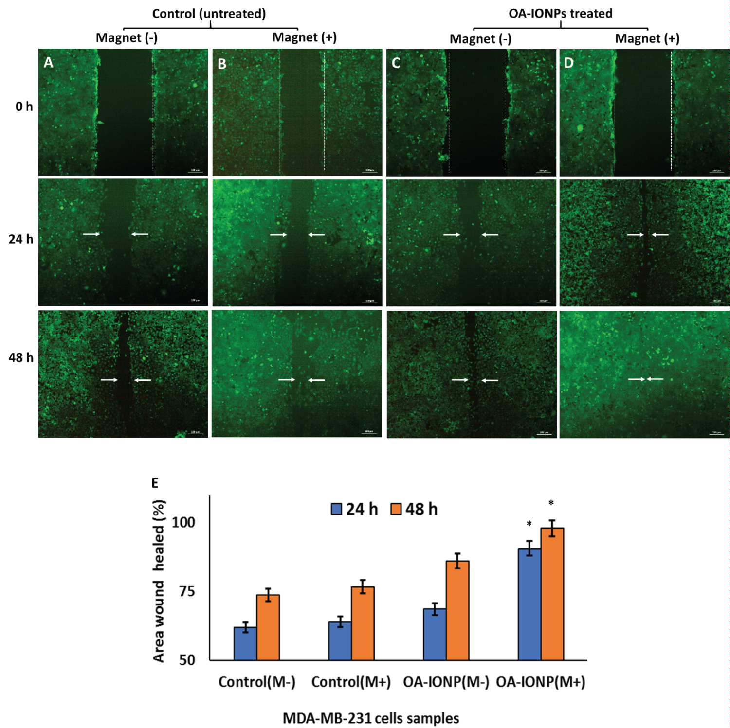
Figure 7: Scratch assay of GFP-tagged...
Scratch assay of GFP-tagged MDA-MB-231 cells preincubated with OA-IONPs (100 µg/mL) showed increased migration of cells under the influence of a magnet. A) Control without magnet; B) Control with magnet; C) OA-IONPs treated cells in the absence of magnet; D) OA-IONPs treated cells in the existence of a magnet, scale bar = 100 µm; E) Represents percentage value of area wound healed calculated using Image J software. Data are expressed as mean ± standard deviation for n = 6 for all four conditions. *p < 0.05 compared to control (untreated) cells.
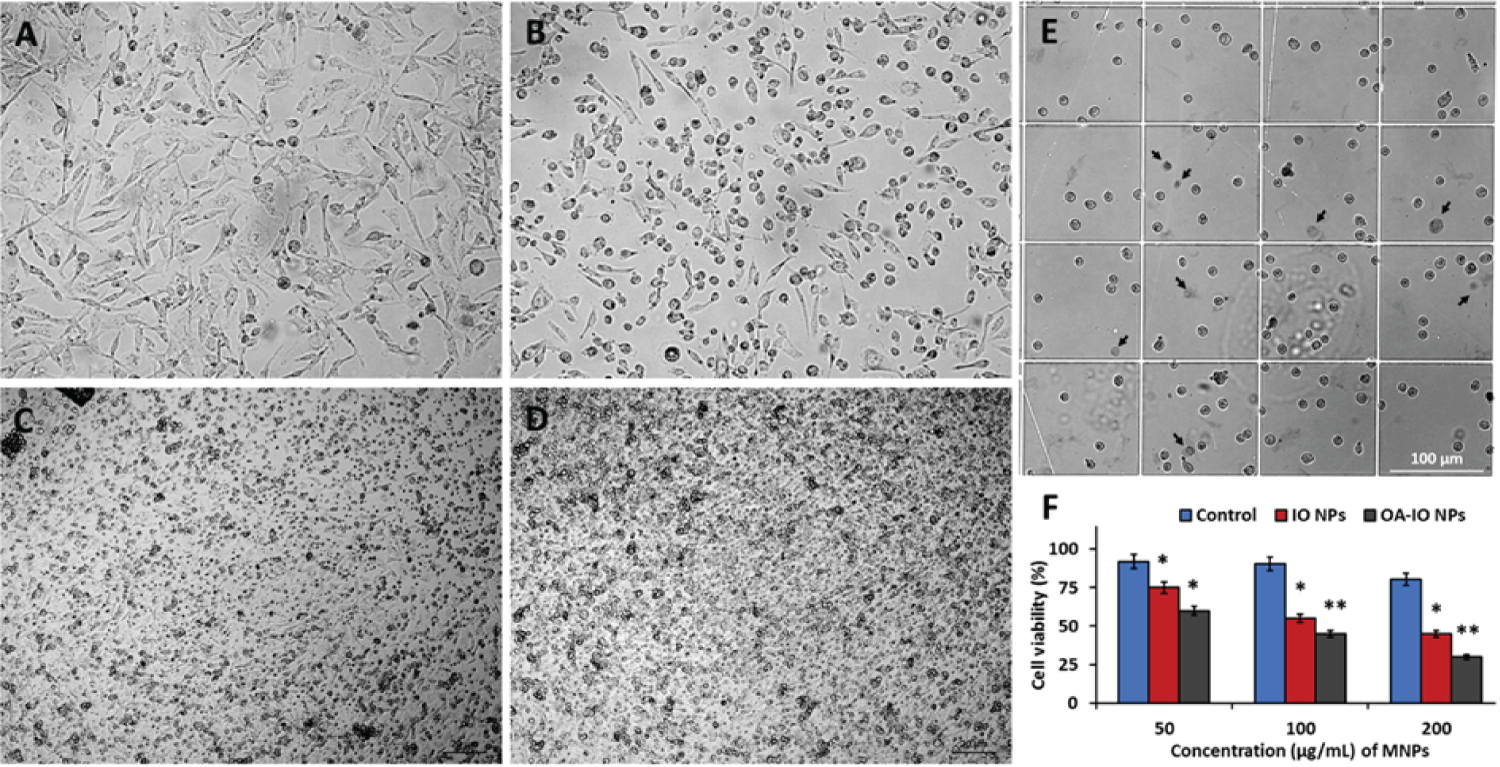
Figure 8: Representative bright-field...
Representative bright-field microscopic images of MDA-MB-231 cells treated with different concentration OA-IONPs and exposed to AMF; A) Control; B) 50 µg/mL; C) 100 µg/mL; D) 200 µg/mL (Scale bar = 20 µm); E) Trypan blue viability staining of MDA-MB-231 cells treated with OA-IONPs at 50 µg/mL concentration, dead cells indicated by black arrows. Scale bar = 100 µm; F) Cell viability analysis by MTT assay of MDA-MB-231 cells after AMF treatment with different concentrations of IONPs and OA-IONPs. Data are expressed as mean ± standard deviation for a minimum of six independent experiments for all concentrations.
*p < 0.05, and, **p < 0.005 compared to control (untreated) cells.
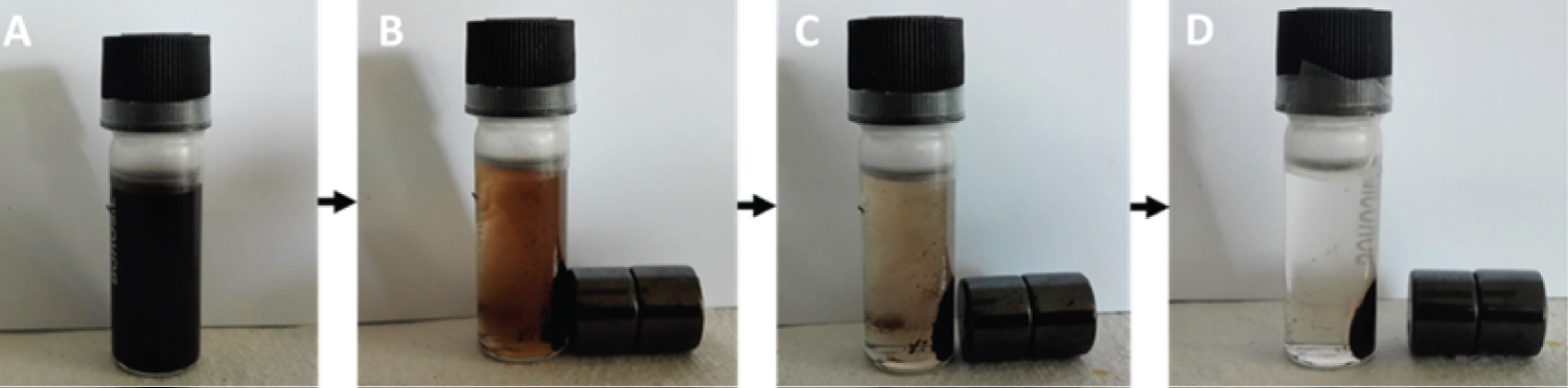
Figure s1: Magnetic separation of...
Magnetic separation of synthesized OA-IONPs suspended in deionised water.
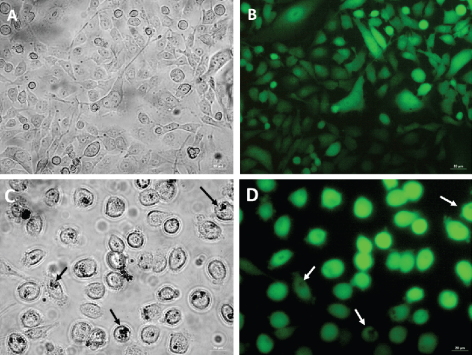
Figure s2: Bright field and fluorescence....
Bright field and fluorescence micrographs of GFP-tagged-MDA-MB-231 cells showed internalization of OA-IONPs; (A) and (B) control, (C) and (D) treated with 100 μg/ml of OA-IONPs. White arrows show the presence of MNPs. Scale bar = 20 μm
References
- Haun JB, Yoon T-J, Lee H, Weissleder R (2010) Magnetic nanoparticle biosensors. Wiley Interdiscip Rev Nanomed Nanobiotechnol 2: 291-304.
- Chen Y, Ding X, Zhang Y, Natalia A, Sun X, et al. (2018) Design and synthesis of magnetic nanoparticles for biomedical diagnostics. Quant Imaging Med Surg 8: 957-970.
- Colombo M, Carregal-Romero S, Casula MF, Gutiérrez L, Morales MP, et al. (2012) Biological applications of magnetic nanoparticles. Chem Soc Rev 41: 4306-4334.
- Mehdaoui B, Meffre A, Lacroix LM, Carrey J, Lachaize S, et al. (2010) Large specific absorption rates in the magnetic hyperthermia properties of metallic iron nanocubes. J Magn Magn Mater 322: L49-52.
- Torti SV, Torti FM (2013) Iron and cancer: More ore to be mined. Nat Rev Cancer 13: 342-355.
- Wang Y, Yu L, Ding J, Chen Y (2018) Iron metabolism in cancer. Int J Mol Sci 20: 95.
- Wu YY, Jiang JN, Fang XD, Ji FJ (2018) STEAP1 regulates tumorigenesis and chemoresistance during peritoneal metastasis of gastric cancer. Front Physiol 9: 1132.
- Hassannia B, Vandenabeele P, Vanden Berghe T (2019) Targeting ferroptosis to iron out cancer. Cancer Cell 35: 830-849.
- Mai TT, Hamaï A, Hienzsch A, Cañeque T, Müller S, et al. (2017) Salinomycin kills cancer stem cells by sequestering iron in lysosomes. Nat Chem 9: 1025-1033.
- Pankhurst QA, Connolly J, Jones SK, Dobson J (2003) Applications of magnetic nanoparticles in biomedicine. J Phys D Appl Phys 36: R167-181.
- Wu W, He Q, Jiang C (2008) Magnetic iron oxide nanoparticles: Synthesis and surface functionalization strategies. Nanoscale Res Lett 3: 397-415.
- Patil RM, Shete PB, Thorat ND, Otari SV, Barick KC, et al. (2014) Non-aqueous to aqueous phase transfer of oleic acid coated iron oxide nanoparticles for hyperthermia application. RSC Adv 4: 4515-4522.
- Yen SK, Padmanabhan P, Selvan ST (2013) Multifunctional iron oxide nanoparticles for diagnostics, therapy and macromolecule delivery. Theranostics 3: 986-1003.
- Nijhawan G, Nijhawan SS, Sethi M (2019) Hyperthermia treatments noble metal-metal oxide hybrid nanoparticles. Elsevier 241-263.
- Wust P, Hildebrandt B, Sreenivasa G, Rau B, Gellermann J, et al. (2002) Hyperthermia in combined treatment of cancer. Lancet Oncol 3: 487-497.
- Ma X, Wang Y, Liu X-L, Ma H, Li G, et al. (2019) Fe 3 O 4 -Pd Janus nanoparticles with amplified dual-mode hyperthermia and enhanced ROS generation for breast cancer treatment. Nanoscale Horizons 4: 1450-1459.
- Tishin A, Shtil A, Pyatakov A, Zverev V (2016) Developing antitumor magnetic hyperthermia: Principles, materials and devices. Recent Pat Anticancer Drug Discov 11: 360-375.
- Gholoobi A, Abnous K, Ramezani M, Homaei Shandiz F, Darroudi M, et al. (2017) Synthesis of γ-Fe 2 O 3 nanoparticles capped with oleic acid and their magnetic characterization. Iran J Sci Technol Trans A Sci 42: 1889-1893.
- Svitkova B, Zavisova V, Nemethova V, Koneracka M, Kretova M, et al. (2021) Differences in surface chemistry of iron oxide nanoparticles result in different routes of internalization. Beilstein J Nanotechnol 12: 270-281.
- Lai CW, Low FW, Tai MF, Abdul Hamid SB (2018) Iron oxide nanoparticles decorated oleic acid for high colloidal stability. Adv Polym Technol 37: 1712-1721.
- Khalil MI (2015) Co-precipitation in aqueous solution synthesis of magnetite nanoparticles using iron(III) salts as precursors. Arab J Chem 8: 279-284.
- Bellova A, Bystrenova E, Koneracka M, Kopcansky P, Valle F, et al. (2010) Effect of Fe 3 O 4 magnetic nanoparticles on lysozyme amyloid aggregation. Nanotechnology 21: 065103.
- Raj R, Das S (2017) Development and application of anticancer fluorescent CdS nanoparticles enriched Lactobacillus bacteria as therapeutic microbots for human breast carcinoma. Appl Microbiol Biotechnol 101: 5439-5451.
- Rawat S, Gupta S, Bhat M, Dinda AK, Mohanty S (2020) Efficient labeling of human Mesenchymal stem cells using iron oxide nanoparticles. Methods Mol Biol 2150: 113-120.
- Yu M, Gai C, Li Z, Ding D, Zheng J, et al. (2019) Targeted exosome-encapsulated erastin induced ferroptosis in triple negative breast cancer cells. Cancer Sci 110: 3173-3182.
- Calatayud MP, Soler E, Torres TE, Campos-Gonzalez E, Junquera C, et al. (2017) Cell damage produced by magnetic fluid hyperthermia on microglial BV2 cells. Sci Rep 7: 8627.
- Aung SM, Kanokwiroon K, Phairatana T, Chatpun S (2019) Live and Dead cells counting from microscopic trypan blue staining images using thresholding and morphological operation techniques. Int J Electr Comput Eng 9: 2460-2468.
- Harris RA, Shumbula PM, Van Der Walt H (2015) Analysis of the interaction of surfactants oleic acid and oleylamine with iron oxide nanoparticles through molecular mechanics modelling. Langmuir 31: 3934-3943.
- Ong HT, Suppiah DD, Muhd Julkapli N (2020) Fatty acid coated iron oxide nanoparticle: Effect on stability, particle size and magnetic properties. Colloids Surfaces A Physicochem Eng Asp 606: 125371.
- Abdullah JAA, Salah Eddine L, Abderrhmane B, Alonso-González M, Guerrero A, et al. (2020) Green synthesis and characterization of iron oxide nanoparticles by pheonix dactylifera leaf extract and evaluation of their antioxidant activity. Sustain Chem Pharm 17: 100280.
- Kershi RM, Ali FM, Sayed MA (2018) Influence of rare earth ion substitutions on the structural, optical, transport, dielectric, and magnetic properties of superparamagnetic iron oxide nanoparticles. J Adv Ceram 7: 218-228.
- Khurshid H, Lampen-Kelley P, Iglesias Ò, Alonso J, Phan M-H, et al. (2015) Spin-glass-like freezing of inner and outer surface layers in hollow γ-Fe 2 O 3 Sci Rep 5: 15054.
- Abenojar EC, Wickramasinghe S, Bas-Concepcion J, Samia ACS (2016) Structural effects on the magnetic hyperthermia properties of iron oxide nanoparticles. Prog Nat Sci Mater Int 26: 440-448.
- Polichetti M, Modestino M, Galluzzi A, Pace S, Iuliano M, et al. (2020) Influence of citric acid and oleic acid coating on the dc magnetic properties of Fe 3 O 4 magnetic nanoparticles. Mater Today Proc 20: 21-24.
- Salwa AA, Karima MZ, Ezzat MS (2020) Facile synthesis of silica coated magnetic nanoparticles via green microwave-solventless technique for purification of water from toxic heavy metals. Int J Nanoparticles Nanotechnol 6.
- Abbasi BA, Iqbal J, Zahra SA, Shahbaz A, Kanwal S, et al. (2020) Bioinspired synthesis and activity characterization of iron oxide nanoparticles made using Rhamnus Triquetra leaf extract. Mater Res Express 6: 1250e7.
- Prates CD, Ballotin FC, Limborço H, Ardisson JD, Lago RM, et al. (2020) Heterogeneous acid catalyst based on sulfated iron ore tailings for oleic acid esterification. Appl Catal A Gen 600: 117624.
- Victory M, Pant RP, Phanjoubam S (2020) Synthesis and characterization of oleic acid coated Fe-Mn ferrite based ferrofluid. Mater Chem Phys 240: 122210.
- Escoda-Torroella M, Moya C, Rodríguez AF, Batlle X, Labarta A (2021) Selective control over the morphology and the oxidation state of iron oxide nanoparticles. Langmuir 37: 35-45.
- Rosales S, Casillas N, Topete A, Cervantes O, González G, et al. (2020) Evaluating physical changes of iron oxide nanoparticles due to surface modification with oleic acid. Chinese Phys B 29: 100502.
- Cai D, Liu L, Han C, Ma X, Qian J, et al. (2019) Cancer cell membrane-coated mesoporous silica loaded with superparamagnetic ferroferric oxide and Paclitaxel for the combination of Chemo/Magnetocaloric therapy on MDA-MB-231 cells. Sci Rep 9: 14475.
- Iacovita C, Fize?an I, Pop A, Scorus L, Dudric R, et al. (2020) In vitro intracellular hyperthermia of iron oxide magnetic nanoparticles, synthesized at high temperature by a polyol process. Pharmaceutics 12: 424.
- Cai J, Miao YQ, Yu BZ, Ma P, Li L, et al. (2017) Large-scale, facile transfer of oleic acid-stabilized iron oxide nanoparticles to the aqueous phase for biological applications. Langmuir 33: 1662-1669.
- Cabrera D, Coene A, Leliaert J, Artés-Ibáñez EJ, Dupré L, et al. (2018) Dynamical magnetic response of iron oxide nanoparticles inside live cells. ACS Nano 12: 2741-2752.
- Augustine R, Alhussain H, Hasan A, Badie Ahmed M, Yalcin CH, et al. (2019) A novel in ovo model to study cancer metastasis using chicken embryos and GFP expressing cancer cells. Bosn J Basic Med Sci 20: 140-148.
- Chernenko T, Matthäus C, Milane L, Quintero L, Amiji M, et al. (2009) Label-free raman spectral imaging of intracellular delivery and degradation of polymeric nanoparticle systems. ACS Nano 3: 3552-3559.
- Schlorf T, Meincke M, Kossel E, Glüer C-C, Jansen O, et al. (2010) Biological properties of iron oxide nanoparticles for cellular and molecular magnetic resonance imaging. Int J Mol Sci 12: 12-23.
- Moaca EA, Farcas C, Coricovac D, Avram S, Mihali CV, et al. (2019) Oleic acid double coated Fe 3 O 4 nanoparticles as anti-melanoma compounds with a complex mechanism of activity- in vitro and in ovo J Biomed Nanotechnol 15: 893-909.
- Fenu M, Bettermann T, Vogl C, Darwish-Miranda N, Schramel J, et al. (2019) A novel magnet-based scratch method for standardisation of wound-healing assays. Sci Rep 9: 12625.
- Zwi-Dantsis L, Wang B, Marijon C, Zonetti S, Ferrini A, et al. (2020) Remote magnetic nanoparticle manipulation enables the dynamic patterning of cardiac tissues. Adv Mater 32: 1904598.
- Hedayati M, Attaluri A, Bordelon D, Goh R, Armour M, et al. (2013) New iron-oxide particles for magnetic nanoparticle hyperthermia: An in-vitro and in-vivo pilot study. Proceedings of SPIE - The International Society for Optical Engineering.
- Ring HL, Gao Z, Sharma A, Han Z, Lee C, et al. (2020) Imaging the distribution of iron oxide nanoparticles in hypothermic perfused tissues. Magn Reson Med 83: 1750-1759.
- Chaparro CIP, Loureiro LR, Valente MA, Videira PA, Borges JP, et al. (2019) Application of hyperthermia for cancer treatment: Synthesis and characterization of magnetic nanoparticles and their internalization on tumor cell lines. 6 th IEEE Port Meet Bioeng (ENBENG), IEEE, Lisbon, Portugal.
Author Details
Ritu Raj1*, Pradipta Ranjan Rauta1, Himanshu Kumar2, Surajit Das3, Aditya Chaubey1, Paul C Salins1, Komal Prasad Chandrachari1 and Bhaskar Viswanathan4
1Mazumdar Shaw Medical Foundation, Bangalore, India
2School of Biotechnology, KIIT Deemed to be University, Bhubaneswar, India
3Laboratory of Environmental microbiology and Ecology (LEnME), Department of Life Science, National Institute of Technology, Rourkela, India
4Department of Radiation Oncology, Vydehi Institute of Medical Science and Research Center, Bangalore, India
Corresponding author
Ritu Raj, Mazumdar Shaw Medical Foundation, Bangalore, India.
Accepted: March 29, 2022 | Published Online: March 31, 2022
Citation: Raj R, Rauta PR, Kumar H, Das S, Chaubey A, et al. (2022) Facile Synthesis of Oleic Acid Capped Iron Oxide Nanoparticles as a Hyperthermic Agent for Cancer Cells Lysis. Int J Nanoparticles Nanotech 7:039.
Copyright: © 2022 Raj R, et al. This This is an open-access article distributed under the terms of the Creative Commons Attribution License, which permits unrestricted use, distribution, and reproduction in any medium, provided the original author and source are credited.
Abstract
The magnetic and thermal properties of iron oxide nanoparticles provide dual advantages as therapeutic and diagnostic agents. Hyperthermia induced by magnetic nanoparticles has emerged as one of the promising cancer modalities either alone or in combinatorial treatment strategies with chemo/radiotherapy in the past few decades. To investigate oleic acid-capped-iron oxide nanoparticles induced cell migration and hyperthermia in MDA-MB-231 cells, a co-precipitation method was used to develop oleic acid-capped-iron oxide nanoparticles from salts of iron by reducing with sodium hydroxide. Human mammary adenocarcinoma cell line, MDA-MB 231 cells were treated with different doses of nanoparticles to study cytotoxicity by MTT assay. Prussian blue staining and invasion assay were used to study the internalization of magnetic nanoparticles by MDA-MB-231 cells. The influence of nanoparticles on cell migration was analyzed by scratch assay, MTT and trypan blue assay were used to analyze cell viability to study magnetic nanoparticle-induced hyperthermia. Synthesis of nanoscale iron oxide particles (< 30 nm) was confirmed by electron microscopy, however, infrared spectroscopy established oleic acid capping over nanoparticles. MTT assay showed 80% of cells remain viable after treatment with high concentrations (200 µg/mL) of iron oxide nanoparticles. The invasion study and Prussian blue assay showed a passive mode of engulfment of magnetic nanoparticles by cancer cells however, the scratch assay has confirmed the influence of magnetic field on cell directional proliferation and migration. Magnetic nanoparticle-mediated hyperthermia showed dose-dependent mortality where 30% of MDA-MB-231 cells survived a dose of 200 µg/mL after exposure to an alternating magnetic field. This study demonstrates the development of oleic acid-capped-iron oxide nanoparticles via co-precipitation and successfully internalization by MDA-MB-231 cancer cells in a time-dependent method. This alternative approach showed an inexpensive mode of targeted annihilation of cancer cells by magnetic nanoparticle-induced hyperthermia.
Keywords
Iron oxide, Nanoparticles, Oleic acid, MDA-MB-231 cells, Magnetic targeting, Cytotoxicity, Hyperthermia
List of Abbreviations
NPs: Nanoparticles; IONPs: Iron Oxide Nanoparticles; OA: Oleic Acid; OA-IONPs: Oleic Acid-Capped Iron Oxide Nanoparticles; MNPs: Magnetic Nanoparticles; GFP: Green Fluorescent Protein; AMF: Alternating Magnetic Field; DLS: Dynamic Light Scattering; FT-IR: Fourier Transform Infrared
Introduction
The biosensing and therapeutic strategies of magnetic nanoparticles (MNPs) have recently received worldwide attention due to their exceptional advantages over other nanosystems. MNPs are physiochemically stable, inexpensive to produce, biocompatible, and environmentally safe [1]. Most of the biological molecules are having insignificant magnetic susceptibilities that made them transparent compared to MNPs under external magnetic fields [2]. The superior magnetic properties of MNPs improves specific absorption rate (SAR) and heating capability as well as radiosensitization of the particles which showed a fast reduction of tumours in the animal model due to highly localized thermal deposition [3,4]. Iron, as a trace element is necessary for cell elementary function and distinctly vital for malignant cells, in which some crucial changes about iron influx and efflux have been recognized. Often, the net outcome of cancer-specific variations is a spike in intracellular iron levels that initiates the activity of iron-dependent proteins and enhances iron proliferation [5]. Usually, iron is attached to transferrin (Tf) glycoprotein in the systemic iron pool, the iron-loaded Tfformulate a complex with transferrin receptor 1 (TfR-1) on the cell membrane and later taken by endocytosis [6]. In the cytosol, Fe 3+ is converted to Fe 2+ by metalloreductases, e.g., six-transmembrane epithelial antigen of prostate (STEAP1-4) family. STEAP1 and STEAP 2 are highly expressed in several human cancer types, e.g., mammary glands, cervix, gastrointestinal organs (colon, pancreas), prostate, bladder, reproductive organs, and Ewing sarcoma [6,7]. The high metabolic activity and proliferation rate of cancer cells are supported by the accumulation of high concentrations of iron and reactive oxygen species (ROS) in these hyperactive cells. However, this can also lead to higher oxidative stress and the activation of cell death pathways. This iron reliance can make cancer cells more susceptible to ferroptosis or iron-catalysed necrosis [8]. The iron oxide nanoparticles (IONPs) can annihilate cancer cells by increasing the concentration of iron and generatingROS. Hydrolysis of IONPs following endocytosis and degradation in the acidic atmosphere of cancer cells can release intracellular iron that increases Fenton-like reactions and ROS generation [9]. IONPs have the potential to serve as both nano-carrier and imaging agents because of their good biocompatibility and spatial imaging capability. They can also generate localized heat when exposed to an alternating magnetic field (AMF), resulting in induced hyperthermia [10-12]. IONPs have shown tremendous potential as a contrast agent in diagnostic radiology and nanomedicine, however, due to their biocompatible and non-toxic nature they have been considered safe for diagnostic and therapeutic purposes by the U.S. Food and Drug Administration (FDA).Multifunctional super paramagnetic iron oxide nanoparticles (SPION) have been used for the treatment of cancer cells by induced hyperthermia, where the functionalized NPs are exposed to AMF to produce localized heat for the annihilation of tumour cells. However, the generation of heat depends upon the size of the particles; small NPs (< 100 nm) heat is produced due to friction between oscillating particles (Brownian mode), and for large NPs (> 100 nm)heat is generated due to the rotation of the magnetic moment with each field of oscillation (Neél mode) [13]. The biological justification for accepting induced hyperthermia is due to the heat sensitivity of tumour cells, which is greater compared to healthy cells. Hence, at high temperatures (41-45 °C) tumour cells get lysed without causing damage to surrounding healthy cells [14]. However, localizing the heat, tumour targeting, and constant temperature distribution across the targeted area are some of the inadequacies of the present methods of delivering induced hyperthermia [15]. Recently, it has been demonstrated that MNPs-induced hyperthermia has an advantage over conventional methods in inducing cell death in vitro . By passive targeting, NPs could be collected at the tumour site and by exposure of AMF, could provide localized heating throughout the tumour [16,17]. On the other hand, large surface-to-volume ratio provides high surface energy to magnetic IONPs and consequently, they tend to aggregate to minimize the surface energy. Additionally, IONPs are being easily oxidized in the air (e.g., magnetite) due to high chemical activity resulting in loss of magnetism and dispersibility. Therefore, it is imperative to offer suitable surface covering and developing effective fortification approaches to retain the stability of magnetic IONPs [18,19]. These approaches include surface-grafting with organic and biocompatible molecules, e.g. surfactants, biopolymers, and biomolecules, or nontoxic inorganic layer (silica, biocompatible metal oxide or metal sulphide). However, in several cases the protective shells stabilize the magnetic IONPs and provide platform for surface functionalization. Oleic acid (OA) is one of the most often used biodegradable capping and stabilizing agents for IONPs due to its capacity to facilitate a dense and protective monolayer around the nanoparticles [20]. IONPs can be efficiently synthesized by co-precipitation method apart from vapour deposition and biological synthesis techniques. As shown below, co-precipitation is the fastest procedure of synthesis and can be easily scaled up even at ambient temperatures [21]. The current study aims at developing a facile method of magnetic IONPs functionalization with biocompatible surfactants; OA for hyperthermia application against invasive cancer.
Experimental
Materials
Ferric chloride (FeCl 3 .6H 2 O, 98%), ferrous chloride (FeCl 2 .4H 2 O, 98%), Oleic acid (OA, ≥ 99%), dimethyl sulfoxide (DMSO) and dimethylthiazolyl-diphenyltetrazolium bromide (MTT) were acquired from Sigma-Aldrich, USA. Sodium hydroxide (NaOH, minimum assay: 98%) was obtained from Chemo Fine Chemicals, India. Dulbecco’s Modified Eagle Medium (DMEM), Fetal bovine serum (FBS), antibiotic (100 IU of penicillin; 100 mg/mL of streptomycin), trypsin, and, phosphate buffer solution (PBS, pH 7.4) were obtained from Thermo Fisher Scientific, USA. All the reagents were of analytical reagent grade and used without any modification/purification. Ultrapure water from a Milli-Q water system (Millipore, USA) was used throughout the study.
Synthesis of Iron oxide nanoparticles
IONPs were synthesized by the co-precipitation method with little modification [22]. Fixed amounts of 0.1 M FeCl 2 .4H 2 O and 0.2 M FeCl 3 .6H 2 O were mixed to make a homogeneous 100 mL solution using deionized water and a magnetic stirrer. The resultant solution was sealed and heated at 60 °C for 15-20 min (using a hot water bath), later mixed with 14 mL of 25% sodium hydroxide (NaOH) solution. After completion of the reaction, the obtained black precipitate was centrifuged at 7000 rpm for 15 min, and washed thrice with deionized water, followed by drying at 60 °C in a hot air oven. For OA-coated IONPs samples, 3 mL of 9.5 mM OA was added into the previously prepared iron mixture and heated at 80 °C. After 1 h, the black precipitate of OA-IONPs was collected from the solution by magnetic separation and washed thrice with deionized water followed by ethanol (99%), later vacuum dried at 60 °C for 12 h.
Characterization of nanoparticles
The physical properties e.g., hydrodynamic diameter, size distribution (Poly Dispersity Index) of MNPs were investigated using dynamic light scattering and zeta potential by Zeta sizer (ZS 90, Malvern Instruments Ltd, Malvern, UK). The lyophilized samples of MNPs were dispersed in sterile PBS to make appropriate colloidal solutions for analysis. These MNPs aliquots were sampled in disposable cuvettes for DLS analysis (diameters, size distribution, and polydispersity index) and zeta potential analysis specialised electrode fitted cells were used. The diameter and size distribution of MNPs were measured at a scattering angle of 90° and 25 °C ambient temperature. Zeta potential and hydrodynamic diameter (size) were measured for each sample in triplicates and their standard deviations were also calculated.
UV-vis absorbance spectra of MNPs samples were analysed in triplicate using UV-Vis spectrophotometer (Multiskan, ThermoFisher, USA) for the wavelength range of 270 nm to 700 nm, where 1 µg MNP sample was dispersed in 1 mL of ultrapure water and sonicated for 5 min before analysis.
The magnetic properties of the synthesized MNPs were analysed by using a vibration sample magnetometer (VSM) under strong magnetic fields (up to 10 kOe). Fourier transform infrared (FT-IR) spectrophotometer (Perkin Elmer, Waltman, MA, USA) was used to examine IR spectra of MNPs over the range of 400-4,000 cm -1 . The morphology of the MNPs was studied using a scanning electron microscope (Nova Nano SEM Field Emission Scanning Electron Microscope, USA) at 20 kV. To increase the electron density of MNPs, synthesized samples were fixed on adequate support and coated with a thin layer of platinum using a platinum sputter module in a high vacuum evaporator. The elemental constituents of the MNPs were analysed by Energy Dispersive X-ray Spectroscopy (EDX, Hitachi, Japan) attached with FESEM. Intricate details of synthesized MNPs were analysed by transmission electron microscope (TEM, JEOL JEM 2100 HR, USA) at 200 kV where MNPs were dispersed in Milli-Q water then cast on a carbon-coated copper grid and air-dried before analysis. The X-ray powder diffraction (XRD) spectrum of prepared nano-formulations was obtained using an X-ray diffractometer (Rigaku Ultima IV, Japan) equipped with a Ni filter and Cu Kα (λ = 1.54056 Å) radiation source. XRD was performed to analyse the crystalline structure of the MNPs.
In vitro studies
The human mammary adenocarcinoma cell line MDA-MB-231 was obtained from Cell Biolabs, Inc., CA, USA (catalogue no. GFPAKR210). The breast cancer cell line was cultured as adherent cells and maintained routinely in DMEM containing 10% FBS and 1% antibiotic solution including penicillin and streptomycin at 37 °C in 5% CO 2 . For viability assessment via MTT assay, cells were seeded onto the 96-well plate and incubated for 24 h before the experiments, and for estimation of live cells in a cell suspension, trypan blue staining was carried using a haemocytometer. For Prussian blue and scratch assay, monolayers of MDA-MB-231 cells were seeded in 6 well plates and 12 well plates respectively.
Cytotoxicity assays
Cytotoxicity effect of OA-IONPs was determined by MTT colorimetric assay after incubation with NPs for 24 h [23]. In brief, MDA-MB-231 cells were seeded at a density of 5 × 10 4 cells/well in a 96-well plate and cultured for 24 h at 37 °C in a 5% CO 2 atmosphere. To the 60-70% confluent cells, the synthesized MNPs of different concentrations (6.25-200 µg/mL) were added in triplicates and further incubated for 24 h. After the incubation period, the media was carefully changed with 100 μL fresh complete media without disturbing cell contents, followed by the addition of 20 μL MTT solution (5 mg/mL). After 4 h incubation at 37 °C, the mixture of DMEM media and MTT solution were removed without disturbing the precipitate (reduced formazan). The obtained precipitate was dissolved in 150 μL DMSO to measure the absorbance of the well contents using a spectrophotometer-based plate reader at 590 nm.
Prussian blue staining
MDA-MB-231 cells treated with OA-IONPs of a fixed concentration were imaged using Prussian blue staining for iron detection [24]. Briefly, control and treated cells were fixed using methanol at - 20 °C for 5 min followed by staining with an equal volume of hydrochloric acid (4%) and potassium ferrocyanide trihydrate (4%) for 15 min and counterstaining with neutral red (0.5%) for 2 min. Prepared samples were then washed with distilled sterile water, air dried, and mounted using dibutylphthalate polystyrene xylene (DPX mountant) medium.
Invasion and scratch assay
Green fluorescent protein (GFP)-tagged-MDA-MB-231 cells were incubated with OA-IONPs for different periods (0, 6, 12, 24 and 48 h). Briefly, GFP-tagged-MDA-MB-231 cells were seeded at a density of 8 × 10 4 cells/well into the 12-well plate and incubated for 24 h at 37 °C in a 5% CO 2 atmosphere. When the cells were 60-70% confluent, the synthesized OA-IONPs of concentration of 100 µg/mL were added in triplicates and incubated for 1 h. The cells were washed with PBS to remove unattached NPs and added with fresh media. Similarly, non-GFP-tagged-MDA-MB-231 cells were incubated with OA-IONPs for 48h and fixed using 3.7% paraformaldehyde. The fixed cells were double-stained with wheat germ agglutinin Alexa Fluor 633 (WGA-Alexa Flour 633) and 4′,6-diamidino-2-phenylindole (DAPI) stain for 15 minutes. The unbounded dyes were removed by washing the cells with PBS. WGA-Alexa stains plasma membrane and DAPI stains cell nucleus. All samples were observed by a fluorescent microscope and images were acquired using ZEISS Axiocam ERc 5s, under acquisition condition set as 358⁄461 nm (excitation/emission) for DAPI and 632/647 nm (excitation/emission) for WGA.
For the scratch assay, the 70-80% confluent GFP-tagged-MDA-MB-231 cells preincubated with OA-IONPs were used, where 1000 μL microtip was used to make a uniform scratch in triplicates [25]. One set of OA-IONPs incubated cells were placed over a small magnet for a period of 48 h and observed by a fluorescent microscope for cell migration.
Image J calculation
For the scratch assay, all the microscopic images were analysed using Image J software for wound area calculation ( https://imagej.nih.gov/ij/ ). Dimension of individual images was calibrated based on scale bar dimension in pixels, and the value of area covered by the wound was measured using the free rectangle selection tool in the Image J software. For every treatment condition minimum of three biological replicates were used and six images per replicate were analysed to obtain average wound area values.
Hyperthermia study
Alternating magnetic field (AMF) experiments were carried out on control MDA-MB-231 cells and MNPs treated MDA-MB-231. The cells were seeded at 1 × 10 6 cells/well and incubated overnight at 37 °C and 5% CO 2 in phenol red-free media with 10% fetal bovine serum and 1% antibiotic solution containing penicillin and streptomycin. The cells were incubated with MNPs at different concentrations (0, 50, 100, and, 200 μg/mL) along with untreated cells as control sample, at 37 °C and 5% CO 2 for 24 h. Afterwards, the cells were washed with PBS and collected using enzymatic dislodging (trypsinization) followed by centrifugation at 7000 rpm for 3 min. The cell pellet of both sets was collected separately and counted using a haemocytometer. Approximately 9 × 10 6 cells were resuspended in 150 μL of DMEM and transferred in a 200 μL PCR tube. The magnetic hyperthermia experiments were performed by placing these PCR tubes in the centre of a thermally insulated copper coil within the magnetic applicator device AMF exposure (f = 560 kHz and H = 23.9 kA/m) for 30 min at the constant target temperature T = 46 °C [26]. For each experiment, the culture plate/PCR tube were equilibrated with the coil for ~15 min before the AMF exposure. Instantly after the field exposure and 4 hours after MNPs treatment, cell viability was measured using trypan blue staining followed by MTT assay, where cells were washed and replenished with fresh growth media and kept at 37 °C and 5% CO 2 for an additional 48 h [27].
Statistical analysis
For the statistical significance of cell viability assays (MTT assays) and scratch assay was assessed by two-way ANOVA in comparison to control (untreated) sets. All data are expressed as mean ± standard deviation for at least six independent experiments for all conditions unless stated otherwise. Statistical analysis was carried out using GraphPad Prism software (GraphPad Software, USA).
Results and Discussion
Characterization of MNPs
The physical observation of colour change (dark black precipitate) and magnetic property of the synthesized MNPs were used for preliminary confirmation of the formation of IONPs with OA coating (Supplementary File 1: Figure S1) [18].
In the present study, DLS, UV-Vis spectrophotometry, FT-IR and VSM have been used to analyse the change in physical properties of IONPs due to OA coating (Figure 1A-E). OA has a binding affinity to surface Fe atoms (Fe 2+ or Fe 3+ ) and forms layer around the NP core which produces a stable structure [28].
DLS, Zeta potential and UV-Vis spectrophotometry
Hydrodynamic diameter and zeta potential of IONPs showed change after coating with OA (Figure 1A and Figure 1B). The OA-IONPs has increased diameter (35.21 nm) and decreased zeta potential (-32.7 mV) compared to 32.12 nm and -25.3 mV of non-coated IONPs respectively. The increase in hydrodynamic diameter of OA-IONPs compared to uncoated IONPs was due to the added thickness of atomic layers owing to a long chain of OA snugging on each other [29].
In UV-vis spectrophotometry, IONPs showed higher absorbance intensity at the range of 300-400 nm wavelength compared to OA-IONPs of similar concentrations (Figure 1C). The absorbance spectra of the MNPs have observed between 342 nm to 374 nm wavelength that corroborated with the earlier reported work [30]. However, due to the electronic transitions of molecules and charge transfer between Fe 2+ and Fe 3+ cations of the synthesized MNPs at their octahedral and tetrahedral sites respectively, the absorbance peaks at UV and near-visible ranges appear prominent. Whereas, a decrease in absorbance intensity of OA-IONPs further confirms OA binding to Fe cations and forming stable OA-capped IONPs [31].
VSM analysis
The magnetic properties of MNPs have shown deviation due to OA coating as was evident in vibrating sample magnetometer (VSM) analysis performed at room temperature (Figure 1D). The saturation magnetization (Ms) 81.03 emu/g at 10 kOe and coercivity of uncoated IONPs were observed 14.69 Oe respectively. The value of Ms has decreased after the surface functionalization of IONPs with OA. Due to the low coercivity and remanence values (less than 10 Oe and 2 emu/g, respectively), the OA-IONPs samples showed significant super paramagnetic behaviour. The loading amount of OA into IONPs could be directly associated with the degree of reduction of Ms from 67.59 emu/g to 52.51 emu/g. The ratio of a disordered spin layer at the surface of IONP to its radius is significantly important however when the size of the resultant particle was decreasing that lead to reduced Ms and reduced saturation magnetization of IONPs [32,33]. Here, the functional layer of OA shields the IONPs from oxidizing and reduces the intra magnetic dipole-dipole interaction between the OA-IONPs. This diamagnetic shell (OA layer) that encapsulated the IONPs might also be credited for lower Ms values compared to noncoated IONPs [34].
FT-IR analysis
FT-IR spectroscopy was used to evaluate the conformational changes of IONPs due to the adsorption of OA. The FT-IR spectra of uncoated IONPs and OA-IONPs showed characteristic peaks at 530, 1046, 1645, and 2975 cm -1 (Figure 1E). However, several additional peaks were detected at 1404, 1451, 1558, 1710, 2856, 2926, and 3290 cm -1 after the surface functionalization of IONPs using OA. The peak that appeared at ~530 cm -1 was allotted to the magnetite phase, however, the peaks at 1645 and 3290 cm -1 were ascribed to the water molecule’s stretching –OH vibrations [35,36]. The supplementary peaks appeared after the OA functionalization of IONPs at 1404 cm -1 was corresponding to the CH 3 molecules of OA, peaks at 1451 and 1558 cm -1 were accredited to the υas (–COO−) and υs (–COO−) stretching respectively, whereas the peaks at 2856 and 2926 cm -1 corresponded to the CH 2 molecules of OA showing asymmetric and symmetric stretching [37,38]. The obtained shift between peaks for υas (–COO−) and υs (–COO−) stretching could be used to determine the interface between the carboxylate ion and the metal (iron) atom [18]. This implies that the appearance of 1451 and 1558 cm -1 peaks in the FT-IR spectrum could be considered as an indication for the formation of superficial OA layer on IONPs [29]. Additionally, the intensity of the peak at 1404 cm -1 was due to the CH 3 triangular mode of OA. The wave number separation of OA-IONPs could be attributed to the covalent interaction between the COO− group and the iron atom as a chelating bidentate [20,39].
Electron microscopy (SEM and TEM), elemental analysis and X-ray diffraction
The anhydrous samples of synthesized MNPs were further scrutinized by electron microscopy. At 15 kV working voltage and 100,000× magnification, spherical homogenous OA-IONPs of average ~12 nm in diameter was observed using FESEM (Figure 2A). TEM analysis of synthesized OA-IONPs was observed at 100 kV over a carbon-coated copper grid without any further adjustment (Figure 2B). Electron microscopy confirmed the formation of nanoscale, homogenous, spherical particles of synthesized OA-IONPs. EDX analysis of various selective areas showed the occurrence of iron in synthesized IONPs along with other residual elements e.g., Cl, Mg, O, and, Si (Figure 2C). The XRD pattern exhibited diffraction peaks at the range of 32°, 36°, 44°, 58°, and, 64° corresponding to(220), (311), (222), (440), and, (331) the characteristics planes of iron oxide (Figure 2D) [39,40].
In vitro studies of the effect of MNPs on MDA MB-231 cells
MDA-MB-231 cancer cells were studied for toxicity, invasion and hyperthermia effect of the MNPs (Figure 3, Figure 4, Figure 5, Figure 6, Figure 7 and Figure 8). MDA-MB-231 cells are adherent and invasive by nature which makes them a suitable experimental choice for studying the effect of therapeutic and diagnostic agents for invasive cancers [41].
Cytotoxicity study by MTT assay
MDA-MB-231 cancer cells were treated with 6.25 - 200 µg/mL of MNPs for time duration of 24, 48 and 72 h (Figure 3A and Figure 3B). As observed, the MNPs did not cause any substantial decrease in cell viability even at a high concentration of 200 µg/mL, however, compared to IONPs, OA-IONPs showed enhanced cytotoxicity. This suggests that the synthesized OA-IONPs were nontoxic at lower dose (≤ 100 µg/mL) however have sublevel of toxicity at higher dosage (200 µg/mL) in case of long time exposure [42,43].
Prussian blue assay for detection of iron in MDA-MB-231 cells
Cells preincubated with OA-IONPs for a fixed-dose (100 µg/mL) were investigated using the Prussian blue staining technique for the presence of iron in cells (Figure 4). The characteristic perinuclear demilune coronas observed surrounding the cell nucleus in MNPs treated cells proved an intracytoplasmic traversing of IONPs [44].
MNPs invasion study in MDA-MB-231 cells
The cellular invasion study of OA-IONPs was performed using green fluorescent protein (GFP) tagged-MDA-MB-231 cells. These cells were treated with 100 µg/mL of OA-IONPs for a duration of 48 h (Figure 5). Similarly, non-GFP-tagged MDA MB231 cells treated with OA-IONPs were observed after 48 h, using dual staining with nucleus (DAPI) and cell membrane marker (WGA 633) at their requisite wavelengths (Figure 6). As it was observed, the cluster of OA-IONPs passively transgresses in the close vicinity of the cell membrane and cytoplasm of MDA-MB-231 cells as they were endocytosed in a time-dependent manner. Between 24 h - 48 h maximum uptake of NPs could be observed, compared to initial hours of incubation. In comparison to the control set, there was no visible cellular irregularity in MNPs treated cells after the incubation period to show any adverse effect of OA-IONPs (Supplementary File 1: Figure S2). The GFP tagged-MDA MB-231 cells are modified cells with a fluorescent cell membrane protein, which can be used for studying intercellular interaction without the application of external fluorescent dyes [45]. The locations of OA-IONPs in the cytoplasm reflect the clustering effects at the point of MNPs transition across the cytoplasm [46]. The sizes of these MNPs clusters represent a great increase in the degree of aggregation concerning the formation of NPs clusters in water, which have hydrodynamic sizes of 100-120 nm for the 35- 42 nm core size nanoparticles [26].
Iron oxide-based NPs have shown a high non-specific phagocytic uptake by tumours and by endothelial cells, where size, charge and concentration of NPs influence their uptake [47]. Seemingly, the charge and coating of the IONPs have a much greater influence on their non-specific uptake than their size. The OA-based coating provides stability and interactive surface for phagocytosis of IONPs by mammalian cells [48]. The phagocytosed NPs reside in the cytoplasm for a very long time before being eliminated from the cells, in our experiment OA-IONPs treated MDA-MB-231 cells showed remnant of IONPs after 3-4 passages.
Cell migration study by scratch assay
The effect of OA-IONPs on cell migration under the influence of magnets was studied using the scratch assay technique. As observed under fluorescence inverted microscope, MNPs treated cells migrated faster under the influence of magnets compared to cells without exposure to a magnetic field. After 24 h, non-treated cells with and without a magnet have covered 63.93% and 61.93% of wound area respectively (Figure 7A and Figure 7B). Cells preincubated with 100 µg/mL of OA-IONPs exposed to a magnet have filled the 90.62% of the wound area but treated cells without a magnet have covered 68.55% of the wound area respectively (Figure 7C and Figure 7D). MNPs treated GFP-tagged MDA-MB-231cells showed preferential migration and healed the wound faster compared to nontreated cells under the effect of external magnetic field [49]. MNPs labelled cells rearranged themselves along the magnetic field direction during the proliferation where intracellular magnetic forces entice the cells to each other while migrating along with the external magnetic fields [50].
Hyperthermia study
The hyperthermia properties of IONPs and OA-IONPs were studies with MDA-MB 231 cells. The cells were incubated with different doses of MNPs 24 h before the exposure to AMF and analyzed by bright field inverted microscope after exposure. The preliminary observation suggested a gradual increase in cellular debris with an increment of MNPs concentration (Figure 8A, Figure 8B, Figure 8C and Figure 8D). After the treatment, cellular viability was assessed by trypan blue assay (Figure 8E) followed by MTT assay (Figure 8F).
The bright-field microscopic images of MDA-MB-231 cells showed an increment in cellular debris which further correlated with cell viability assay of the same. At 50 µg/mL around ~60% of cells survive after being exposed to AMF, however, it decreased to ~30% with a 200 µg/mL concentration of OA-IONPs. It was established experimentally that the induced magnetic hyperthermia effect of IONPs improves after functionalization with OA.
The hyperthermia properties of OA-IONPs demonstrated therapeutic relevant heating at low AMF amplitudes in an in-vitro set-up and could be easily scaled for in-vivo tests [51]. The possible reason for the greater hyperthermic effect is the functional OA layer which can retain the superparamagnetic fraction of the IONPs. The surface layer of OA also inhibits the aggregation of IONPs in the aqueous phase and forms a stable suspension of OA-IONPs as compared to noncoated IONPs [52]. It is reported earlier that the stable colloidal solution of superparamagnetic NPs enhances the hyperthermic effect through Brownian movement and Neel's spin relaxations [12,42,53]. Thus, the hyperthermia study strongly supports that the functionalization of IONPs with OA has a beneficial effect on its therapeutic value.
Conclusion
The successful synthesis of OA-IONPs was accomplished through co-precipitation of salts of Fe 2+ and Fe 3+ ions under alkaline conditions. These particles with an average diameter of < 30 nm formed a stable colloidal solution in aqueous media. Further, the cell viability assay suggests that the MNPs were nontoxic at concentrations below 200 µg/mL. Thus, these particles at lower doses lower doses can be used for further studies on hyperthermia generation under magnetic induction. The physicochemical characterizations revealed properties of IONPs e.g. size, shape, surface charge, functional groups, magnetic properties etc. However, in-vitro studies e.g., cellular uptake study, magnetic hyperthermia studies etc. are being intended to evaluate the feasibility of the nanosystems in vitro and in vivo for hyperthermia applications, especially for invasive tumour/cancer cells. The thermal ablation properties of MNPs are directly related to the size and dimensionality of NPs. IONPs have strong hyperthermia action which could be explored for in-vivo experiments. In summary, these studies have established that in the intricate world of MNPs, therapeutic ablation using an effective chemically improved magnetic hyperthermia treatment for malignant tumours in physiologically acceptable magnetic fields could be feasible.
Consent for Publication
All authors agreed for publishing the manuscript in its present form.
Availability of Data and Materials
All data generated or analysed during this study are included in the article.
Funding
This work was financially supported by Vision Group for Science & Technology, Government of Karnataka (CESEM, GRD No. 180).
Conflict of Interest
The authors declare no conflict of interest.
Acknowledgement
The authors acknowledge Mazumdar Shaw Medical Foundation, Narayana Hrudayalaya, NH city, Bangalore for providing equipment and resources and National Institute of Technology, Rourkela for helping in the characterization of nanoparticles.