
International Journal of Robotic Engineering
(ISSN: 2631-5106)
Volume 3, Issue 1
Review Article
DOI: 10.35840/2631-5106/4107

Article Formats
Automatic Image and Video Analysis for Capsule Endoscopy - An Open Frontier
Table of Content
References
- Iddan G, Meron G, Glukhovsky A, Swain F (2000) Wireless capsule endoscopy. Nature 405: 417.
- VBS Prasath (2015) On fuzzification of color spaces for medical decision support in video capsule endoscopy. Modern Artificial Intelligence and Cognitive Science (MAICS) 147-151.
- VBS Prasath, R Delhibabu (2014) Automatic contrast enhancement for wireless capsule endoscopy videos with spectral optimal contrast-tone mapping. International Conference on Computational Intelligence in Data Mining (ICCIDM), Springer SIST 31: 243-250.
- VBS Prasath, R Delhibabu (2015) Automatic mucosa detection in video capsule endoscopy with adaptive thresholding. Computational Intelligence in Data Mining (ICCIDM), Bhubaneswar, India, Springer SIST 410: 95-102.
- VBS Prasath, R Delhibabu (2014) Automatic image segmentation for video capsule endoscopy. International Conference on Computational Intelligence: Health and Disease (CIHD), Visakhapatnam, India. Proc. Springer CIMI 73-80.
- VBS Prasath, H Kawanaka (2015) Vascularization features for polyp localization in capsule endoscopy. IEEE International Conference on Bioinformatics and Biomedicine (BIBM), Washington, DC, USA. Proc. IEEE 1740-1742.
- VBS Prasath (2017) Polyp detection and segmentation from video capsule endoscopy: A review. J Imaging 3: 1.
- VBS Prasath (2017) Automatic bleeding detection from video capsule endoscopy-A review, Submitted, 2017. Arxiv.
- VBS Prasath, H Kawanaka (2017) Near-light perspective shape from shading for 3D visualizations in endoscopy systems. IEEE International Conference on Bioinformatics and Biomedicine (BIBM), USA, IEEE, 2293-2295.
Author Details
VB Surya Prasath1,2,3,4*
1Division of Biomedical Informatics, Cincinnati Children's Hospital Medical Center (CCHMC), USA
2Department of Biomedical Informatics, College of Medicine, University of Cincinnati, USA
3Department of Electrical Engineering & Computer Science, University of Missouri-Columbia, USA
4Department of Electrical Engineering & Computer Science, University of Cincinnati, USA
Corresponding author
VB Surya Prasath, Department of Electrical Engineering & Computer Science, University of Cincinnati, OH 45221, USA.
Accepted: June 28, 2018 | Published Online: June 30, 2018
Citation: Prasath VBS (2018) Automatic Image and Video Analysis for Capsule Endoscopy - An Open Frontier. Int J Robot Eng 3:007.
Copyright: © 2018 Prasath VBS. This is an open-access article distributed under the terms of the Creative Commons Attribution License, which permits unrestricted use, distribution, and reproduction in any medium, provided the original author and source are credited.
Abstract
Imaging the gastrointestinal (GI) tract is challenging due to the tortuous nature of colon and many of the traditional colonoscopy based imaging systems could not reach all the regions. Video capsule endoscopy (VCE) is a revolutionary imaging system wherein tiny cameras packed into a pill-sized capsule obtain images without any tethered wires and transmit the data using wireless technology. This has enabled the gastroenterologists to obtain almost painless endoscopy exams of patients with full visual coverage of the colon. However, VCE exams obtain video data that is composed of a large number of frames, thus require tedious manual viewing and analysis to identify possible GI tract anomalies. Automatic image and video analysis techniques are sorely lacking in this area and we provide a focused review of our developed solutions along with the challenges that remain to be solved in capsule endoscopy image/video processing and analysis.
Keywords
Capsule endoscopy, Image analysis, Video processing, Automatic inspection, Computer-aided diagnosis
Introduction
Capsule endoscopy is an imaging technique that provides visualizations of the gastrointestinal (GI) tract [1]. It opened the door for visualizations of hard to reach regions of the GI tract that are not captured by traditional colonoscopy systems. Video capsule endoscopy (VCE) is a wireless imaging system in a capsule form that contains cameras capturing continuous imaging data (color images) of the GI tract and transmits them to a signal receiver worn on the patient's waist. The capsule moves through the GI tract without any external forces and uses only the natural peristalsis motion. This unhindered motion means there is little or no discomfort to the patient, and the capsule exits naturally. Typically the video data consists of around 55000 frames in color (RGB data [2]), and are analyzed in a workstation by gastroenterologists for possible abnormalities.
Despite these imaging advantages, the sheer number of images gastroenterologists need to examine poses great challenges, as it requires careful attention and tedious manual processing of all the image frames per each VCE exams. Also, human operators can miss subtle abnormalities that can be captured by automatic image analysis methods that can discern at pixel, patches, and features levels. Despite the availability of mature automatic image and video processing techniques that work for natural images, and other medical imaging modalities with good performance/accuracy, there aren't many reliable automatic algorithms available for VCE imagery. After the introduction of VCE in 2000's there have been some automatic computer-aided diagnosis (CAD) systems that are designed to analyze the VCE data. In this review, we focus on some of the automatic image and video analysis methods developed by us for processing VCE images, and further challenges that remain to be solved.
Advances in Image/Video Processing for VCE
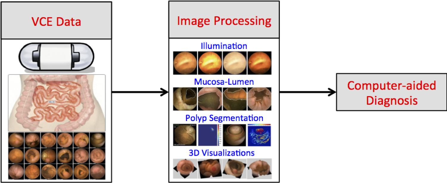
Similar to any other medical imaging modality VCE requires a well-defined sequence of image processing/analysis tools from low to high-level algorithms to obtain a fully developed CAD pipeline. Figure 1 shows a typical VCE image processing, and analysis pipeline that consist of the following important components.
• Illumination estimation/correction
VCE contains multiple near-lights, that is, the camera and the surrounding LED lights are situated closer to the surface (mucosa) that is being imaged. Thus, inhomogeneous light saturation is visible and requires robust illumination preprocessing to avoid missing important mucosa tissue characteristics [3].
• Mucosa-lumen separation
Separating mucosal tissue and lumen (the area in which the capsule moves) regions is another important preprocessing step required to characterize features on mucosa rather than the lumen [4,5].
• Polyp segmentation/localization
Polyps can be precursors to GI cancer, thus identifying and detecting them is an important image processing problem. However given the large amount of frames each VCE exam, this requires robust polyp indicator functions [6] and efficient algorithms [7].
• Bleeding segmentation/detection
GI bleeding require a careful analysis of the color information and machine learning algorithms that are trained on prior examples are essential in obtaining good detection results [8].
• 3D visualizations
Three dimensional visualizations of the GI tract can provide further insight into the GI tract [9].
A final CAD system that consists of these off-line image analysis tools can be of assistance to gastroenterologists in making decisions based on VCE images.
Challenges
Automatic abnormalities detection without manual supervision is perhaps one of the most important open challenges in this area. Despite the advancements in some of the low level image processing tasks such as illumination correction, mucosa-lumen separation, the higher-level tasks such as polyp segmentation/localization, bleeding segmentation/detection require further research. Moreover, automatic video processing methods that exploit the temporal information are required to obtain well-defined CAD systems.